Pathologies and Treatments

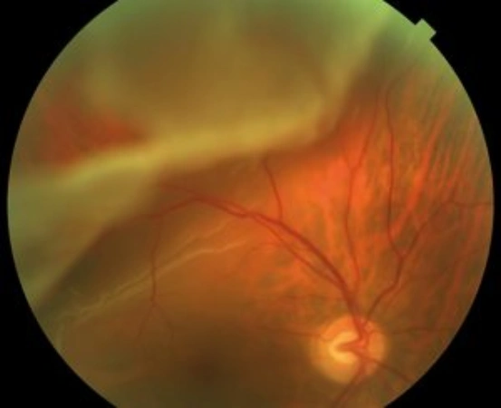
Retinal detachment has an incidence of approximately one in every 15,000 people. This event, which can occur at any age, is more frequent in myopic patients or those with a history of the disease.

The retina is a thin layer that covers the inner part of the posterior wall of the eye, where the images that are sent to the brain are focused. Under normal conditions, the retina is attached to a more external layer, the retinal pigment epithelium. Conceptually, retinal detachment refers to the separation between the layers of the neurosensory retina and the retinal pigment epithelium.

Symptoms

The main symptom is usually the perception of repetitive flashes of light in a sector of the visual field. This symptom would indicate a retinal tear, a condition that can lead to retinal detachment. Other symptoms may include partial or total loss of the visual field, perceived as a black curtain blocking vision.

Early detection of a retinal tear and subsequent treatment with laser photocoagulation can prevent retinal detachment.

Treatments

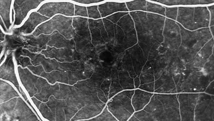
A thorough ophthalmologic examination is essential, including a complete study of the peripheral retina to diagnose all retinal tears responsible for retinal detachment. Sometimes, optical coherence tomography and ocular ultrasound help to make the diagnosis.

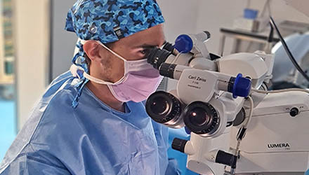
Treatment is almost always surgical and consists of reattaching the retina. The goal is to reposition the retina and create adhesion between the neurosensory retina and the retinal pigment epithelium by sealing the retinal tears with laser photocoagulation.

There are different surgical techniques for the treatment of rhegmatogenous retinal detachment, such as vitrectomy, scleral surgery, and pneumatic retinopexy. For a correct choice, each case must be analyzed and individualized.

The Institut de la Màcula has the most advanced technology for the treatment of retinal detachment. Choosing the best surgical technique in each case allows us to address the treatment of this pathology with the highest guarantees of success.

Medicina regenerativa: Endoret

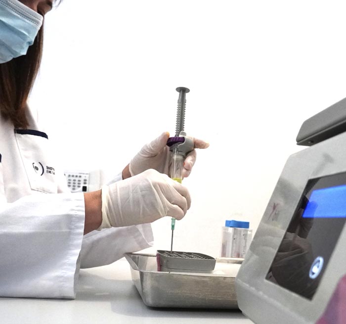
Con el tratamiento de medicina regenerativa Endoret propiciamos la reparación de la superficie ocular afectada por la sequedad ocular. Se trata de un plasma rico en factores de crecimiento para el tratamiento de las lesiones de la superficie ocular elaborado a partir de la sangre del mismo paciente. Con esta formulación individualizada se consigue una tolerancia completa y una efectividad óptima. Esta opción terapéutica revolucionaria es muy eficaz en casos de defectos epiteliales persistentes o úlceras corneales. Asimismo se puede aplicar después de una cirugía refractiva corneal, una cirugía ocular o en el Síndrome de Sjögren.

Procedures

Medicina regenerativa: Endoret

Con el tratamiento de medicina regenerativa Endoret propiciamos la reparación de la superficie ocular afectada por la sequedad ocular. Se trata de un plasma rico en factores de crecimiento para el tratamiento de las lesiones de la superficie ocular elaborado a partir de la sangre del mismo paciente. Con esta formulación individualizada se consigue una tolerancia completa y una efectividad óptima. Esta opción terapéutica revolucionaria es muy eficaz en casos de defectos epiteliales persistentes o úlceras corneales. Asimismo se puede aplicar después de una cirugía refractiva corneal, una cirugía ocular o en el Síndrome de Sjögren.

Dr. Jordi Monés,

M.D. PhD

Número de Col·legiat COMB: 22.838

Director. Doctor of Medicine and Surgery. Specialist in Ophthalmology. Specialist in Retina, Macula, and Vitreous.

If you found this interesting, feel free to share it here:

Last modified: 01/09/2025
Scroll to Top
Privacy Overview

This website uses cookies so that we can provide you with the best user experience possible. Cookie information is stored in your browser and performs functions such as recognising you when you return to our website and helping our team to understand which sections of the website you find most interesting and useful.