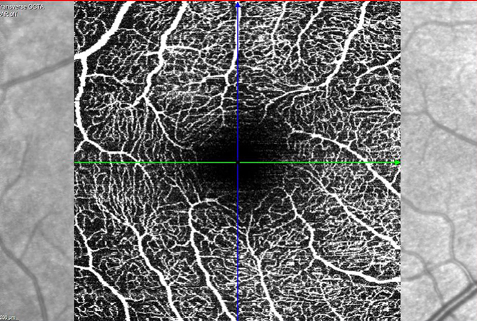
OCT angiography (OCT-A) is a new, fast, and non-invasive imaging modality that allows evaluation of the retinal microvascular network. OCT-A can provide a three-dimensional image of the retinal microvasculature and detect capillary abnormalities in patients with diabetic retinopathy and other retinal vascular diseases.
With OCT-A, the superficial vascular plexus and the deep capillary plexus can be examined separately, which is not possible with conventional techniques such as fluorescein angiography or indocyanine green angiography. Therefore, OCT-A provides additional information.
OCT-A is performed similarly to optical coherence tomography (OCT), but unlike conventional angiography, OCT-A does not require intravenous contrast injection.