Pathologies and Treatments

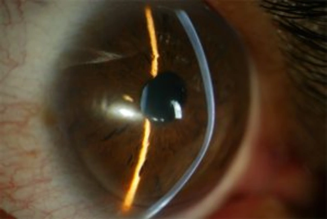
Keratoconus is the most common non-inflammatory axial corneal ectasia. It is characterized by central or inferior paracentral thinning, which causes a progressive increase in corneal curvature. This results in myopia, irregular astigmatism, and conical apical protrusion.

Keratoconus is the leading cause of corneal transplantation in young patients. The etiology is not fully known and is likely multifactorial. A positive family history has been described in 6%–8% of cases. It is likely that if corneal topography were performed on relatives of affected patients, the incidence would be higher. In other cases, it appears sporadically, linked to complex inheritance patterns that are still under investigation.

There are also cases related to ocular allergy or continuous eye rubbing. The typical form of presentation occurs at puberty and progresses over the following ten to twenty years. The rate of progression tends to be higher the younger the patient is.

Symptoms

In the early stages, the patient may have good visual acuity, even without correction, and the only evident sign is the difficulty in obtaining an accurate refraction.
In the intermediate stages, the patient presents decreased vision and a sudden increase in astigmatism, which cannot be fully corrected with glasses but improves with the use of rigid or gas-permeable contact lenses. Due to the progression of astigmatism, the patient requires frequent changes of glasses and contact lenses.
When keratoconus is very advanced, visual acuity is impaired both with glasses and contact lenses due to irregular astigmatism, an increase in higher-order aberrations, and corneal scarring.
Astigmatism, unlike myopia and hyperopia, does not usually evolve naturally with age. For this reason, if a sudden increase in astigmatism is detected in children or young patients, it is important to visit the ophthalmologist to rule out keratoconus.
It is very important to diagnose the disease in these patients at early stages, because with treatments to slow its progression, the prognosis of keratoconus has greatly improved, and the need for corneal transplants has decreased.

Treatments

There are several treatment options depending on the progression of keratoconus.
Certain cases of keratoconus are mild enough, at least for some time, to allow vision to be adequately corrected with glasses. However, rigid or gas-permeable contact lenses are more useful in all but mild cases.
In cases where progression and evolution of keratoconus are documented, corneal cross-linking has proven to be very effective in halting its progression.
Intrastromal rings regularize the cornea by correcting the deformation caused by keratoconus.
In more advanced cases, when vision has significantly decreased or when central corneal scarring is detected, the indicated treatment is corneal transplantation, which can now be performed by selectively replacing only the affected layers of the cornea. This is called lamellar keratoplasty.

Medicina regenerativa: Endoret

Con el tratamiento de medicina regenerativa Endoret propiciamos la reparación de la superficie ocular afectada por la sequedad ocular. Se trata de un plasma rico en factores de crecimiento para el tratamiento de las lesiones de la superficie ocular elaborado a partir de la sangre del mismo paciente. Con esta formulación individualizada se consigue una tolerancia completa y una efectividad óptima. Esta opción terapéutica revolucionaria es muy eficaz en casos de defectos epiteliales persistentes o úlceras corneales. Asimismo se puede aplicar después de una cirugía refractiva corneal, una cirugía ocular o en el Síndrome de Sjögren.

Procedures

Medicina regenerativa: Endoret

Con el tratamiento de medicina regenerativa Endoret propiciamos la reparación de la superficie ocular afectada por la sequedad ocular. Se trata de un plasma rico en factores de crecimiento para el tratamiento de las lesiones de la superficie ocular elaborado a partir de la sangre del mismo paciente. Con esta formulación individualizada se consigue una tolerancia completa y una efectividad óptima. Esta opción terapéutica revolucionaria es muy eficaz en casos de defectos epiteliales persistentes o úlceras corneales. Asimismo se puede aplicar después de una cirugía refractiva corneal, una cirugía ocular o en el Síndrome de Sjögren.

Dra. Paula Verdaguer,

MD, PhD

Número de Col·legiat COMB: 40737

Ophthalmologist. Specialist in Cornea, Refractive Surgery, and Cataracts

If you found this interesting, feel free to share it here:

Last modified: 26/08/2025
Scroll to Top
Privacy Overview

This website uses cookies so that we can provide you with the best user experience possible. Cookie information is stored in your browser and performs functions such as recognising you when you return to our website and helping our team to understand which sections of the website you find most interesting and useful.