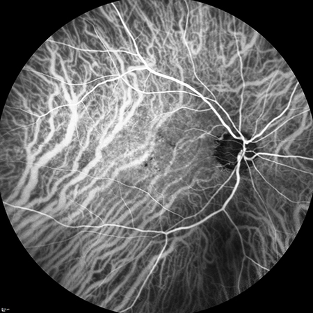
Indocyanine green angiography is a diagnostic procedure that uses a specific dye (indocyanine green) to detect leaks or damage in the blood vessels that supply the retina. Indocyanine green exhibits its characteristic staining properties when exposed to invisible infrared rays. Performing this examination requires sophisticated cameras sensitive to these light wavelengths.
This angiography is used to study the deep vessels of the choroidal layer. It is particularly valuable in the treatment of age-related macular degeneration (AMD), as indocyanine green can distinguish the deep choroidal vessels, especially those hidden or blocked by blood. It is especially useful for detecting variants of exudative AMD, such as polypoidal choroidal vasculopathy, which has important therapeutic implications.
It also helps identify feeding neovessels of neovascular membranes, which in certain cases can be directly treated through photocoagulation, reducing or avoiding the need for intravitreal injections.
The procedure is similar to standard angiography: the patient is injected with indocyanine green dye, which circulates through the bloodstream, while photographs are taken using a specialized camera.