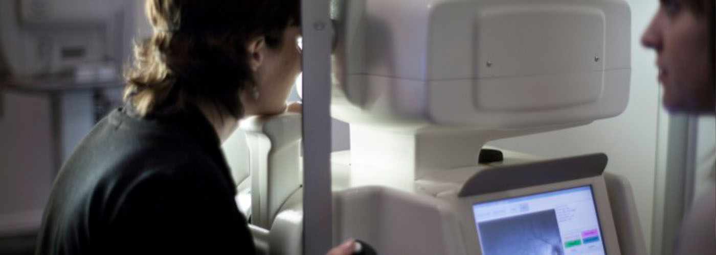
Microperimetry is one of the most specific tests to determine central vision defects and the extent of damage caused by certain diseases such as age-related macular degeneration, diabetic retinopathy, diabetic macular edema, and central serous retinopathy.
Microperimetry is a non-invasive technique that maps the macular visual field. It is performed first on one eye and then on the other, and does not require pupil dilation. It currently represents a new tool to assess visual function in a wide range of retinal disorders. The microperimeter is considered the ideal instrument for this examination, with advantages related to its ability to perform automatic tracking that ensures a rapid and precise analysis of retinal sensitivity, fixation location, and stability. Several studies have shown that the results of this test correlate significantly with those provided by optical coherence tomography (OCT) in retinal diseases, enabling comprehensive evaluation of human visual function through the morphology-function relationship.


