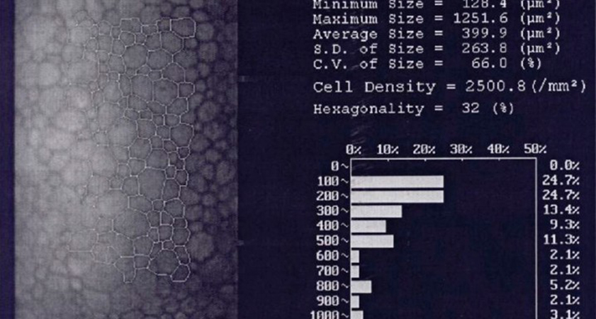
Specular microscopy allows us to study the corneal endothelium. Since the cellular distribution is nearly uniform, this technique determines the number, shape, and size of the endothelial cell population.
Specular microscopy provides a reflected image of the optical interface between the corneal endothelium and the aqueous humor, but it can also be used to obtain images of the epithelium, corneal stroma, and lens.
A normal endothelium in a young person shows, on specular microscopy, a regular pattern of hexagonal cells, most with similar diameters, which can vary due to age, trauma, pathologies, or surgical procedures.
The procedure is non-contact. Eye alignment is achieved using fixation light, and it is recommended that the patient blink several times before image capture to moisten the ocular surface and enhance brightness. Specular microscopy is an essential non-invasive diagnostic aid that assesses the condition and physiological reserve of the endothelial cell layer, considering that a clear cornea does not guarantee a normal corneal endothelium. Therefore, we systematically perform this test on patients before cataract surgery and phakic intraocular lens implantation surgery.