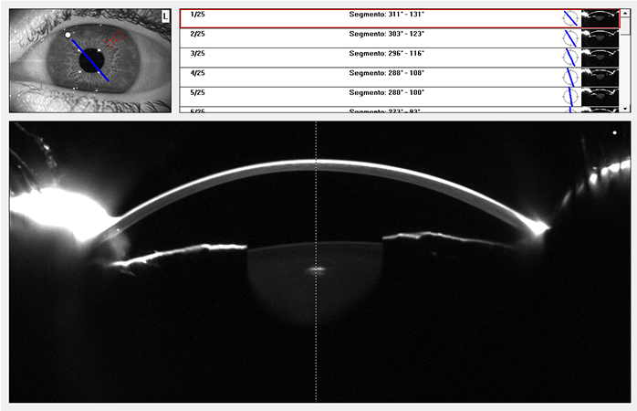
Optical coherence tomography (OCT) allows us to obtain high-resolution cross-sectional images of the anterior and posterior segments of the eye non-invasively, using optical interferometry. It has become a very useful tool for the ultrastructural study of ocular anatomy.
Anterior segment OCT is used to monitor patients who have undergone refractive surgery, intracorneal ring implants, corneal cross-linking, corneal transplants, phakic intraocular lenses, and filtering glaucoma surgery. In cataract surgery, OCT enables precise analysis of incision architecture and the relationships between the intraocular lens and the posterior capsule.
Anterior segment OCT is very useful for analyzing and evaluating tumors and cysts of the anterior segment, conjunctival tumors, and various corneal conditions such as dystrophies, degenerations, or infections. It also allows determination of corneal and epithelial pachymetry, quantification of the iridocorneal angle, measurement of anterior chamber depth, evaluation of positioning and alignment of phakic and pseudophakic intraocular lenses, study of contact lenses on the cornea and the ocular surface status, and is helpful in the study of dry eye syndrome.


