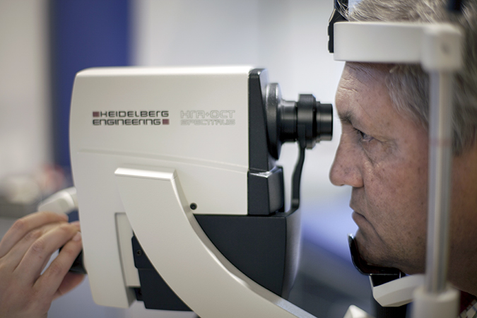
Optical coherence tomography (OCT) is a non-invasive optical tomographic imaging technique (sectional imaging) that uses a combination of light sources from various detectors to achieve higher resolution. The millimetric penetration of this technique provides high-resolution images of the different retinal layers. The latest generation technology available at the Institut de la Màcula allows identification of abnormal details at an almost cellular scale in different retinal layers and multiple sections scanning the entire macular area, enhancing diagnostic capability and monitoring of various therapies for macular and retinal pathologies. Today, managing macular pathology without the diagnostic aid of optical coherence tomography would be inconceivable.
Optic Nerve OCT
Optical coherence tomography (OCT) of the optic nerve (ON) provides quantitative measurement of the pupil and retinal nerve fiber layer, which is very useful for diagnosing and monitoring patients with glaucoma.
Measuring the retinal nerve fiber layer helps differentiate between healthy eyes and those affected by glaucoma. Furthermore, by comparing results between examinations, it allows detection of glaucoma progression.


