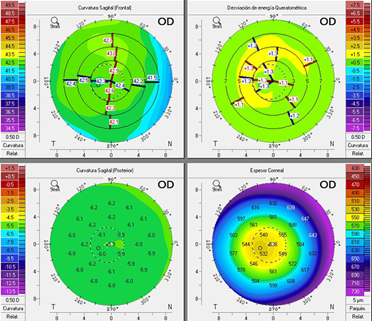
Corneal topography performs a systematic analysis of the cornea, allowing a thorough understanding of its shape.
Modern topographers incorporate projection systems (Placido discs) and elevation measurements to provide a more complete analysis of the anterior and posterior corneal surfaces.
Currently, one of the most important applications of corneal topography is the early detection and monitoring of corneal ectasia. The combined data analysis enables reliable diagnosis, follow-up, and determination of the most effective therapeutic strategy based on the magnitude and type of astigmatism (regular or irregular).
However, corneal topography is also essential before corneal refractive surgery, in refractive cataract surgery to calculate the power of the intraocular lens to be implanted, in keratoplasties, and in corneal abnormalities, among other uses.
No single value alone holds definitive weight in diagnosing a pathology; it is important to analyze all parameters provided and their clinical correlation.
No special preparation is required for this exam. The patient only needs to have a well-hydrated ocular surface. Contact lens wearers should discontinue use for 10–15 days before the exam to avoid affecting the results.


